
Common skin conditions — burns, pimples and the like — are diagnosed by looking closely, which a webcam cannot do. The digital microscope at a ClinicWala e-clinic captures magnified, well-lit images and video of the skin for the doctor on the consultation.
It plugs into a desktop, PC, smartphone or tablet over USB, so the image can be shared and analysed the moment it is taken — and captured photos and video track how a condition changes between visits.
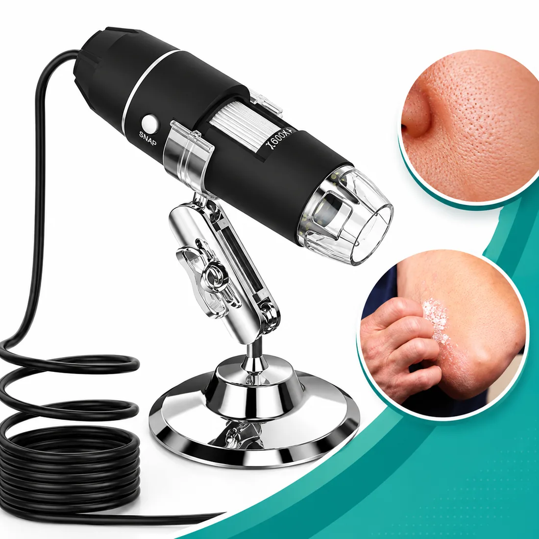
What the device does
Plugs into anything
A USB interface connects the microscope to desktops, PCs, smartphones and tablets — the image is on a screen and shareable immediately.
40x to 1600x magnification
A focus wheel selects the magnification, from an overview at 40x to fine detail at 1600x.
Eight adjustable LED lights
Built-in LEDs with adjustable brightness and focus length keep the image clear and well lit, even in a dim room.
Flexible metal stand
An adjustable stand holds the microscope steady at any angle and height, so photos and video come out sharp.
Plug and play
Powered over the USB cable, no charging needed, and the bundled software works out of the box.
Real-time capture
Still images and video are captured live, so a skin condition can be tracked from one visit to the next.
The numbers behind the device
- Magnification
- Up to 1600X
- Video pixels
- 640*480P
- Imaging range
- Manual adjustment
- Interface
- USB 2.0
- Power supply
- USB (5V DC)
- Adaptive frames
- Up to 30 fps at 600 LUX
Certifications
The manufacturer states the following certifications for this device: CE.
Ask the franchise team for current certificates when you evaluate the kit.