
A fundus camera photographs the retina — the light-sensitive layer at the back of the eye where conditions like glaucoma, macular degeneration and diabetic retinopathy first show. A doctor on a video call cannot look into your eye; a clear fundus photograph lets an eye specialist do exactly that, remotely.
The camera at a ClinicWala e-clinic is compact and lightweight, supports both mydriatic and non-mydriatic imaging, and works with PCs, smartphones and tablets. Images appear in real time during the examination and go into your ClinicWala health record for the specialist reading them.
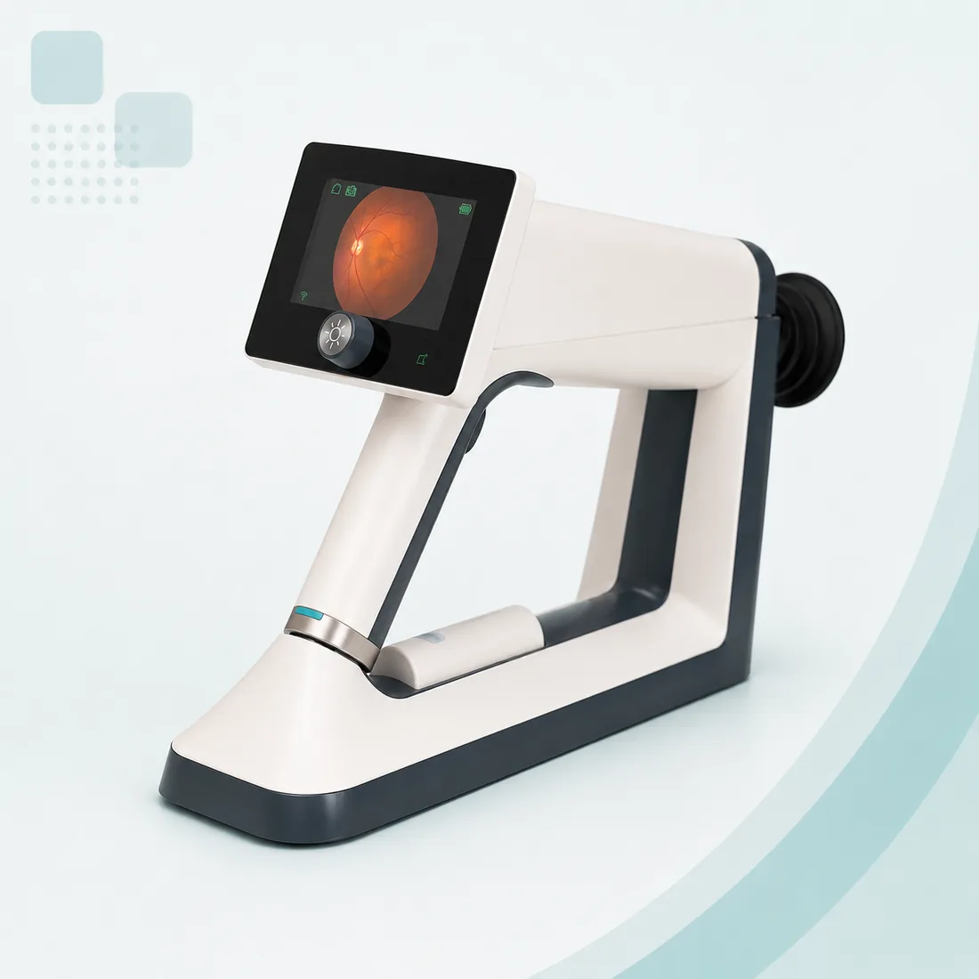
Where the e-clinic uses it
Eye screening
Detailed retinal imaging and fundus photography support early detection and monitoring of eye diseases such as glaucoma, macular degeneration and diabetic retinopathy.
Telemedicine consultations
Clear, detailed images during a video consultation let a remote ophthalmologist examine the eye properly — more accurate diagnoses for eye complaints.
Diabetic retinopathy screening
For diabetic patients, regular high-quality retinal photographs catch retinopathy early — while treatment can still protect vision.
What the device does
High-resolution imaging
Captures the fine retinal detail a medical assessment depends on.
Real-time imaging
The image appears as it is captured, giving immediate visual feedback during the examination and the consultation.
Simple to operate
A straightforward interface any e-clinic operator can use — no imaging specialist needed at the counter.
Works with everyday devices
Compatible with PCs, smartphones and tablets, so images move easily into the consultation and the record.
Portable and lightweight
Compact enough to carry and use across clinical settings, not fixed to one room.
The numbers behind the device
- Imaging modalities
- Mydriatic and non-mydriatic
- Light source
- Infrared and cool white LED
- Dioptre correction
- -30D to +30D
- Fixation target
- Internal, 8 points
- Minimum pupil size
- 3mm
- Working distance
- 33 mm
- Field of view
- 40 degrees
- Battery
- Rechargeable; up to 7 hours non-mydriatic, up to 3 hours mydriatic
- Capture modes
- Manual and auto
Certifications
The manufacturer states the following certifications for this device: CE, FDA, ISO.
Ask the franchise team for current certificates when you evaluate the kit.